Portfolio
Exocytosis & endocytosis
An animation showing vesicles fusing to a cellular membrane. This was a personal project with the aim of highlighting the merging and displacement of lipids when fusion occurs (highlighted with the various colours of lipids).
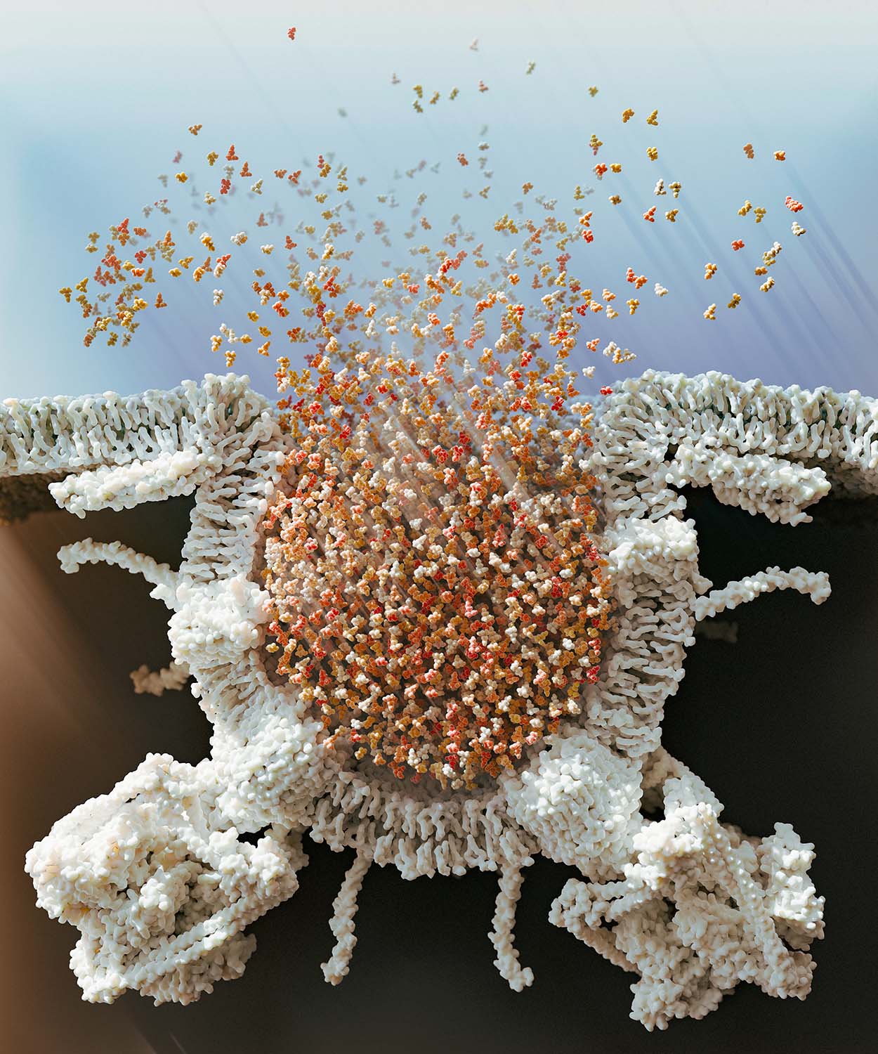
Synaptic popcorn
Synaptic vesicles are absolutely packed with glutamate - around 8000 molecules on average - this concentration is depicted here in a 40nm diameter vesicle. Also shown are some of the main vesicle proteins present in their reported average abundance, such as V-ATPase, glutamate transporters and SNARE complexes.
Cytonemes in the germarium niche
How cytonemes influence signalling in the germarium niche.

Cell division
A short physics-based cell division animation. The animation was not made to portray a particular process, but in my mind it is reminiscent of embryo development, spheroid growth of suspended cells in culture, or tumour growth.
Ligand binding to receptor
Depicted are two cells, one with Notch receptors (red), and the other with Delta ligands (blue). This short animation uses data from the Protein Data Bank to show an accurate final binding position of the receptor and ligand. The membrane is procedurally animated.
Wellcome Trust Cell-Matrix Centre 30th anniversary posters
This set of posters was designed for the Wellcome Trust Cell-Matrix Centre's 30th anniversary event that took place at The University of Manchester on the 25th June 2025. The Cell-Matrix centre is comprised of 30 research groups that broadly work on three distinct research areas: Mechano-matrix, Immuno-matrix, and Chrono-matrix. Therefore the idea behind the posters was to highlight these three areas in a visually compelling way while also providing more technical depth by the inclusion of graphical abstracts describing a selection of the research that has been carried out by the groups at the centre.

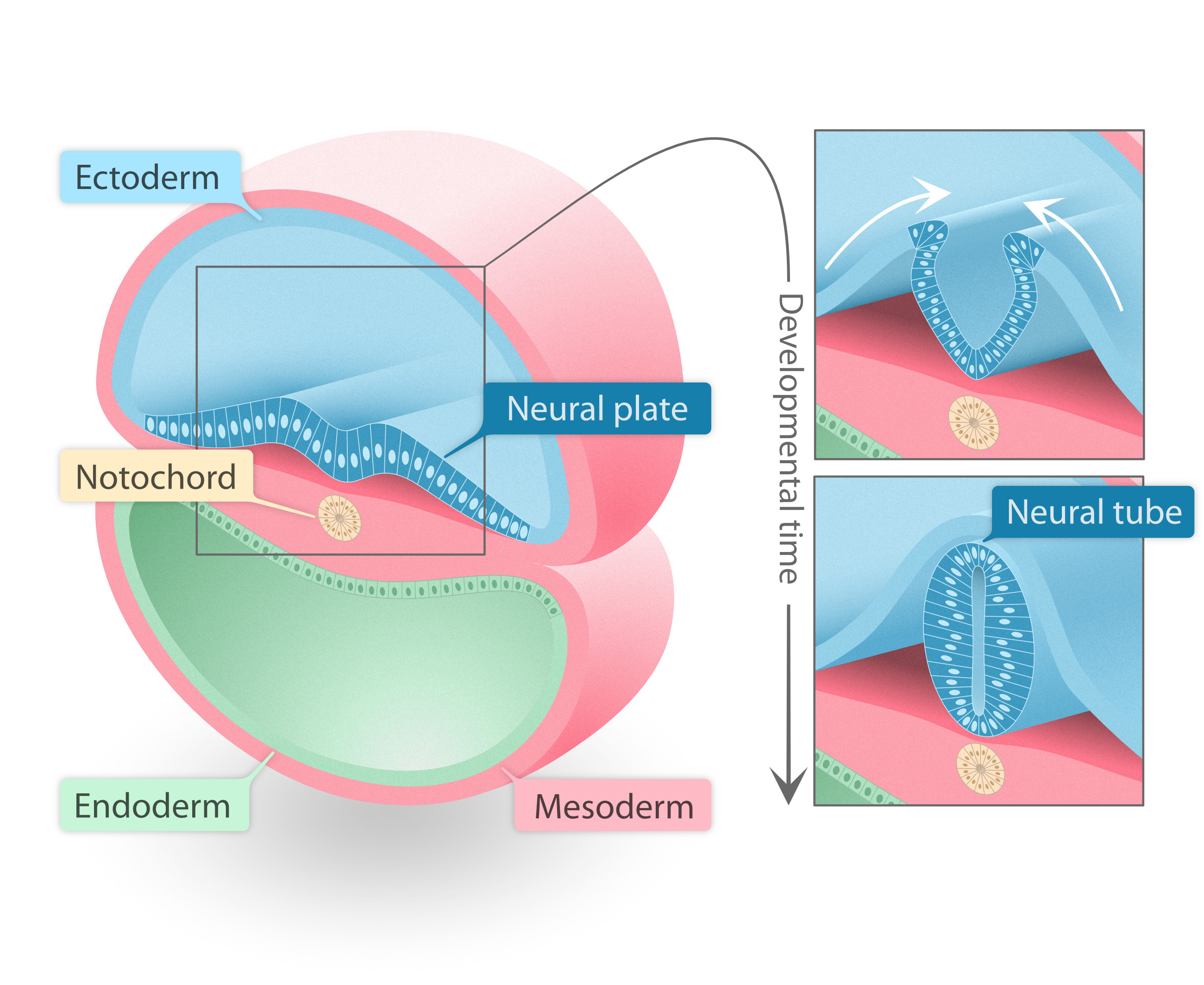
Neurulation: the developing spinal cord
The process of neurulation. The ectoderm forms the spinal cord by first folding in on itself to form the neural tube, which is made up of neural progenitors that go on to form the neurons and glia of the spinal cord, brain, and retina.
Interkinetic nuclear migration in neuroepithelia
A procedural animation of neuroepithelia, with a focus on visualising interkinetic nuclear migration.
Potassium ion channel
A procedural animation of a phospholipid bilayer, potassium ion channels and potassium ions passing through them.
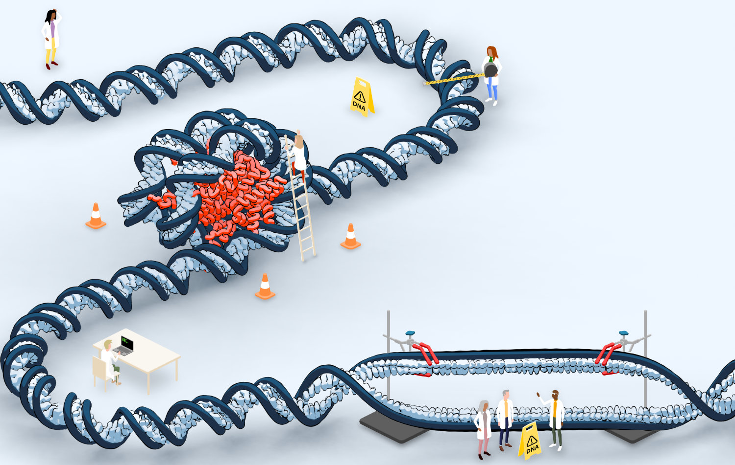
The PhD process
Concept art for a PhD recruitment advert. This image depicts the journey of taking on a scientific research area during a PhD.
BMP signalling
Paper figure depicting modifications to BMP signalling.

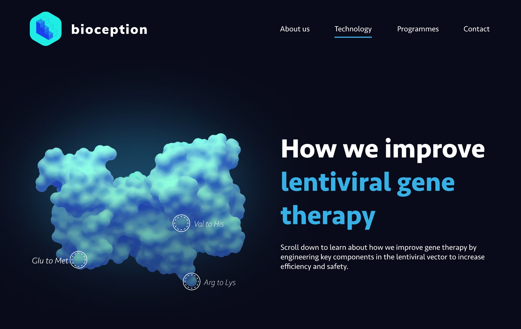
Webpage banner
Webpage banner designed for the Translation Toolkit at The University of Manchester. Link to Translation Toolkit.

UK In-Situ Resource Utilisation logo
A logo developed for the UK In-Situ Resource Utilisation (ISRU UK) group who are funded by the UK Space Agency. They represent research in the UK that concerns space exploration resource utilisation.